Causes and risk factors
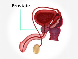
Retention of urine can be caused either due to obstructive or nonobstructive causes. Nonobstructive causes can be neurogenic (weak bladder muscle or nerves), traumatic due to certain medications, any tumors of the urinary system, enlargement of the prostrate (in males) along with calculi in kidney, urethra, or bladder are the major obstructive factors hampering the flow of urine. Nonobstructive causes can also lead to the same. Retention of urine occurs due to weak pelvic floor structures or due to weak urethral sphincter or bladder muscle. It is commonly seen in women after delivery due to damage to the pelvic floor structures. Trauma or injury to the internal structures during road traffic accidents can also lead to the same. In men, high incidence is seen due to prostate enlargement. It is also seen in athletes who are engaged in heavy exercises. Overactivity of detursor muscles at old age or detursor muscle abnormalities can also lead to retention. Diseases like spina bifida, multiple sclerosis, spinal cord injury affects the function of the nerve which supply the bladder. Urinary infection, severe constipation, and side effects of certain medications are other contributing factors.
Clinical presentation:
Retention of urine is of two types – acute and chronic. In acute type, the bladder is full but there is an inability to evacuate it. It occurs suddenly and can be life threatening. There is inability to pass urine. Complete obstruction is seen. Pain and discomfort along with bloated feeling is felt in the lower abdomen.
In chronic urinary retention, the person can urinate but the bladder is not completely emptied. There is increased frequency to pass urine. There is difficulty in initiating the urine and difficulty while passing it. The flow is weak and dribbling is seen. The urine has a foul odor. There is a sense of pressure in the lower abdomen.
Investigations:
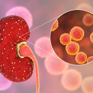
Symptoms narrated by the patient and the physical examination carried out by the doctor will help to confirm the diagnosis. Postvoid urine measurement is done. Urodynamic studies are advised. Certain investigations which are done are routine blood tests, urine analysis, ultrasonography of the pelvis, and abdomen. Cystogram and cystoscopy are other valuable investigations which can be done.
Treatment:
In cases where there exists some underlying disease, treating the same is the most essential step. Appropriate conservative and surgical methods need to be adopted. Bladder drainage is done. It consists of draining the urine using a catheter.
Certain pelvic floor exercises, i.e., Kegel exercises to strengthen the pelvic muscles are advised. These are exercises which cause repeated contraction and relaxation of the muscles of the pelvic floor. It is done with the help of a pelvic toner device. In cases which do not respond well to behavioral training and exercises, medications are prescribed. Use of absorbent pads or certain devices like pessary or fixer-occluder device is advised. In cases where incontinence is caused due to diseases like prolapse of pelvic organs or weak sphincter, it needs to be corrected by surgery. Other surgical procedures like midurethral sling, bulking around urethra are adopted. In some cases, electrical nerve stimulation is done.
When to contact a doctor:
Seek an advice from a doctor if one suffers from any urinary complaint, especially difficulty in passing urine and/or pain or discomfort in lower abdomen.
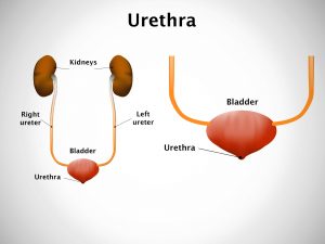
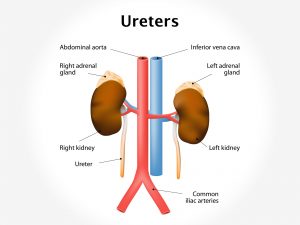
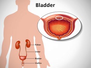
System Involved: Urinary system
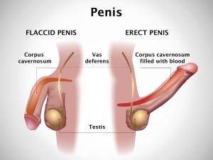
An organ involved: kidney, bladder, urethra, nerves, and muscles