Causes and risk factors
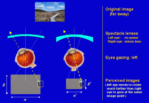
Aniseikonia may be present naturally or it may be induced. Induced aniseikonia can be optically induced or retinally induced. Optically induced aniseikonia can occur by the use of corrective spectacles or surgery to treat refractive errors. Retinally induced aniseikonia occurs as a result of tearing or detachment of retina, macular edema, or the presence of epiretinal membranes.
Clinical presentation
Objects viewed from different eyes and those viewed bilaterally appear of varying sizes. This can cause the brain to subconsciously suppress one of the eyes. The patient may develop headache due to eyestrain. Other symptoms include dizziness and loss of balance.
Investigations
Aniseikonia can be demonstrated by an eye examination. The patient is asked to observe an object placed directly in front of one eye at a distance of about 15 cm. The patient is asked to observe the object from each of his eyes separately and then with both eyes together. On doing this, the object will appear larger to the eye that it is directly in front of.
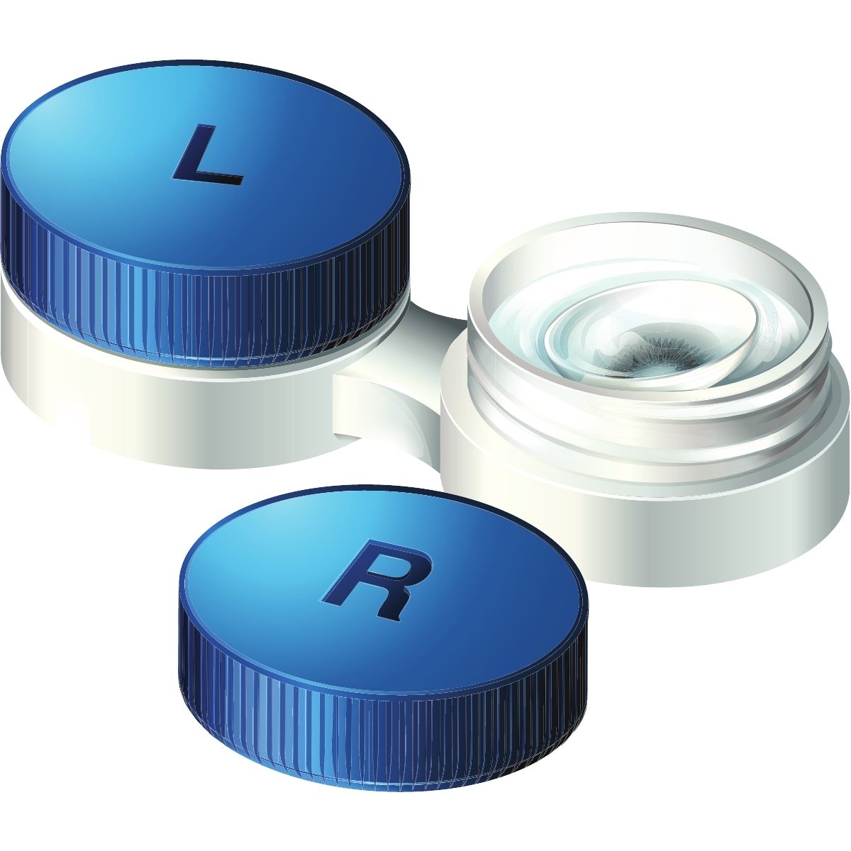
Treatment
Corrective spectacles and lenses are used to correct the refractive errors. The degree of correction can be determined by the ‘aniseikonia value.’ The ‘aniseikonia value’ is a measure of how much correction is needed in the right eye in order to normalize the vision. For e.g., an aniseikonia value of -10% suggests that the right eye is perceiving objects about 10% larger than their actual size. Hence minification of images by 10% is necessary in order to normalize the vision.
When to contact a doctor
Contact a doctor as soon as you experience any eye symptoms such as diplopia or disproportionate object sizes.
Systems involved
Ophthalmology.
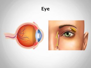
Organs involved
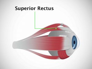
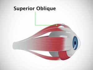
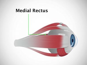
Eyes.