Causes and risk factors
Normally the arteries carry the blood away from the heart while the veins carry the blood to the heart. Capillaries are small blood vessels which connects the veins and arteries.
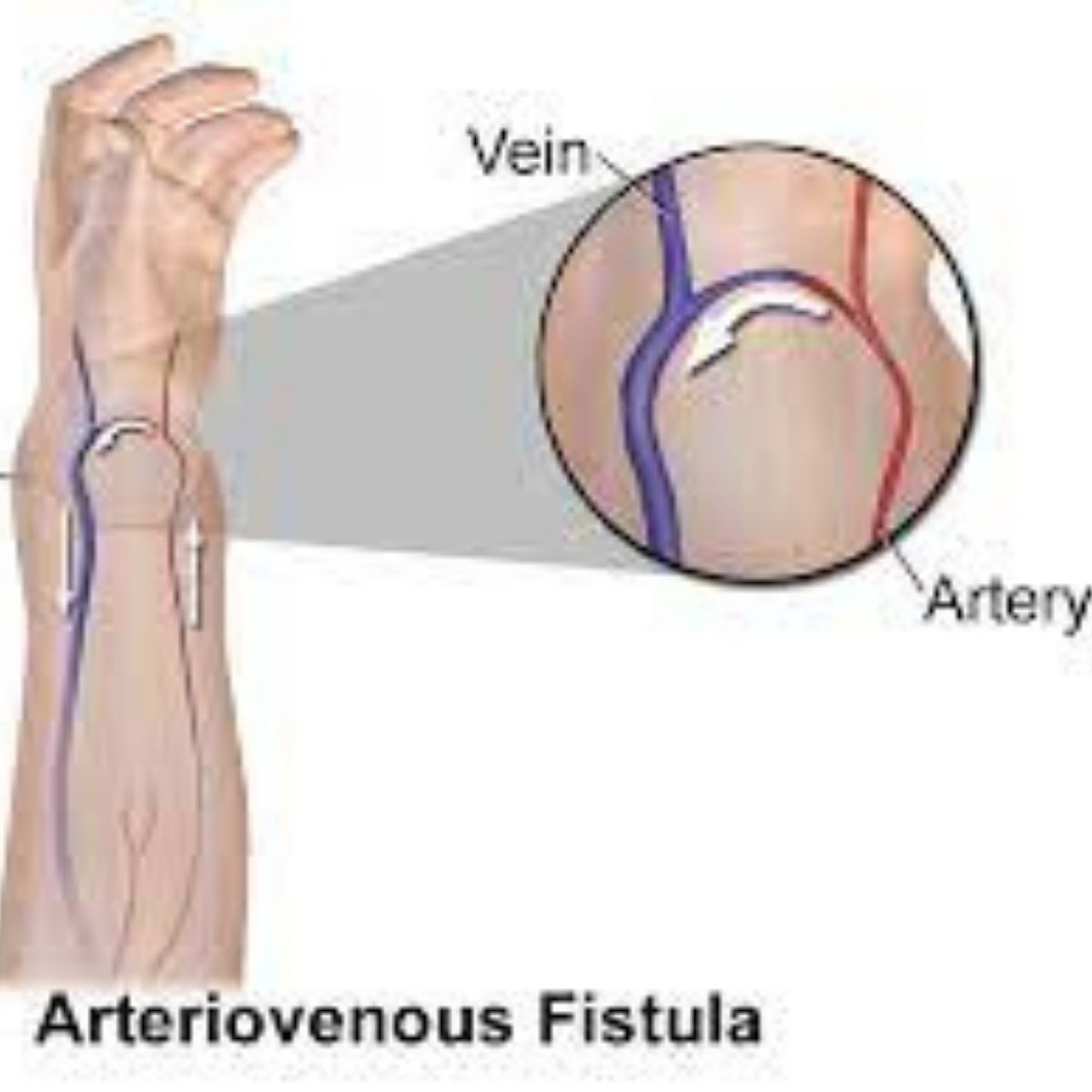
In arteriovenous fistula the blood from the artery directly enters the veins. An arteriovenous fistula can be present since birth or it may be developed later in life. Penetrating injury to the artery or vein is the most common cause of arteriovenous fistula. Arteriovenous fistulas can also be made artificially by a vascular surgeon for treatment purpose e.g.: During hemodylasis.
Clinical presentation:
Congenital arteriovenous fistula near the surface of the skin, gives a skin a reddish blue appearance as if bruised. The affected area is swollen. Fall in blood pressure results in overworking of the heart causing palpitation. On auscultation of the site murmur can be heard. Over time this overload on heart can lead to heart failure. Arteriovenous fistulas can lead to potentially life threatening diseases like stroke, hemorrhages or formation of blood clots. Artificial arteriovenous fistulas are usually made by a vascular surgeon. Preferably forearm or arm is the site of choice. This AV fistula provides extra flow of blood with extra pressure giving a vascular access. It is a blood flow during hemodylasis in end stage renal diseases.
Investigations:
Diagnosis is done of the basis of the symptoms narrated by the patient and the physical examination carried out by the doctor. Doppler Ultrsonography, CT Scan or MRI are diagnostic tools. Certain other routine investigations can also be recommended by the doctor.
Treatment:
Monitoring is required in cases where the fistula is small and does not pose any health issues. Arteriovenous fistulas are surgically repaired either by open surgery where the damaged part is cut off or laser coagulation therapy is adopted. While the above treatment is adopted as a first line of treatment in some diseases, an artificial AV fistula is recommended in certain therapeutic procedures.
