Causes and risk factors
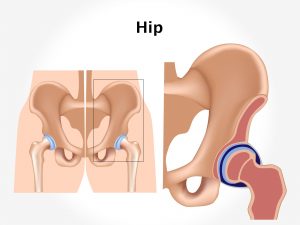
Congenital dysplasia of hip is common in first born children. During first pregnancy, the abdominal muscles of the mother is tight and it causes restriction of fetal movement. Breech presentation is an important responsible factor. Genetic factor also plays an important role in its causation. An individual who has a family history of dysplasia of the hip increases the risk 30 times. As the incidence is high in girls, it suggests that maternal gonadotropins stimulate the production of progesterone in female fetus. This causes increased laxity of joints. Congenital dysplasia of the hip is commonly associated with neuromuscular abnormalities like cerebral palsy, spina bifida arthrogryposis, etc.
Clinical presentation:
This condition is diagnosed while conducting physical examination in newborn babies. During infancy the mother may complain of unequal opening of the leg while changing the nappy. Shortening of femur may be noticed. Unequal limb length can be seen. In congenital dysplasia, creases are seen on the thigh skin. As the child grows, walking becomes difficult, limping is seen in kids. Waddling gait is seen in cases where bilateral affection is seen.
Investigations:
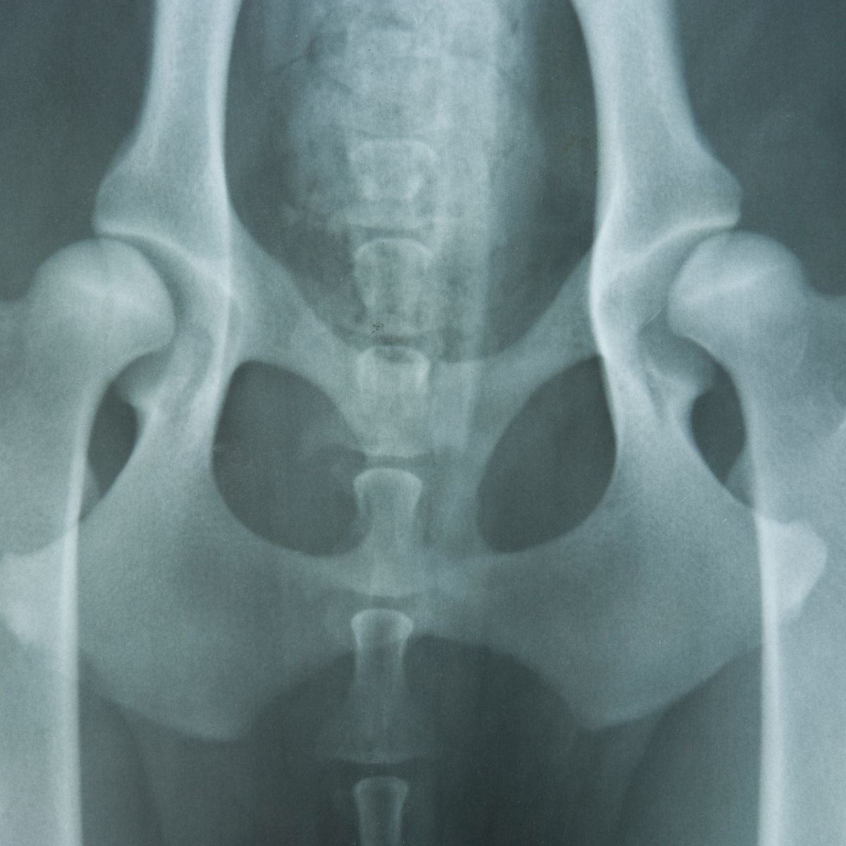
The symptoms narrated by the mother or the physical examination carried out by the doctor helps in confirming the diagnosis. In newborn babies, certain physical tests like the Ortolani test and Barlow test can be done. A plain x-ray is usually sufficient for diagnosis. Certain other tests – basic and specialized along with certain imaging tests can also be advised.
Treatment:
The treatment plan depends upon the age of the patient. In cases of presence of any underlying cause, it needs to be treated. In neonates, use of splint or harness is done. The use of harness, particularly the Pavlik harness, is more commonly used. Surgical intervention is advised in patients between the age of 6 months to 2 years. Closed reduction technique is used. If this is not possible, then open reduction along with realignment procedures are done. Plaster spica is applied for immobilization for about 6 months. In children above the age of 2 years, the treatment is usually difficult as secondary changes have already taken place. Femoral osteotomy is carried out. For improving the strength and activity of the muscles, exercises are advised.
Other Modes of treatment:
Certain other modes of treatment can also be helpful in coping up with the symptoms. Taking into consideration the symptoms in a holistic way, homoeopathy can offer a good aid for the relief of the symptoms. The Ayurvedic system of medicine which uses herbs and synthetic derivates can also be beneficial in combating the complaints. Certain yoga exercises can also be helpful in strengthening the muscles.
Facts and Figures:
The incidence is seen in 20 per 1000 live births.
