Causes and risk factors
Diaphragm serves as an important muscle during ventilation. Diaphragmatic paralysis is idiopathic in most cases. Paralysis can be of 2 types – single leaflet involvement, i.e., unilateral diaphragmatic paralysis [UDP] or both leaflet involvement, i.e., bilateral diaphragmatic paralysis [BDP]. Although diaphragm mostly performs work of respiration, there is a role of accessory respiratory muscles in ventilation. In case of BDP, all of the work is performed by accessory muscles. Thus, eventually they fatigue which leads to ventilation problems. Causes include peripheral phrenic nerve injuries, motor neuron disease, neuropathies, and myopathies. Conditions like stroke, viral infections, tumors, trauma, and inflammation can cause diaphragmatic paralysis. Subdiaphragmatic processes and operations can also lead to the disorder. Rare causes include systemic lupus erythematosus, neck trauma, central vein cannulation, and occult thoracic malignancy. The condition may occur following liver transplantation.
Clinical presentation
Symptoms depend on whether the paralysis is unilateral or bilateral. Unilateral includes asymptomatic presentation, whereas diaphragmatic paralysis is incidentally discovered in patients going for chest radiography for some other reason. Patients usually are asymptomatic at rest, but experience dyspnea on exertion and have a decrease in exercise performance. If the patient has an underlying lung disease, dyspnea may occur at rest. Orthopnea may be present. Bilateral presentation includes dyspnea that worsens in the supine position, tachypnea and rapid, shallow breathing occurs when the patient adopts the recumbent position. Patients also report anxiety, insomnia, morning headache, excessive daytime fatigue, and poor sleep habits. Respiratory failure may occur. In some patients, nonspecific GI symptoms such as heartburn, regurgitation, nausea, and epigastric pain can also develop.
Investigation
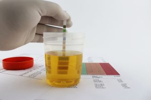
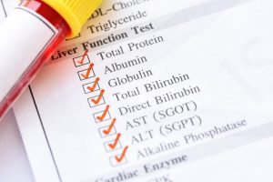
Medical history by the patient and clinical examination by the doctor helps in diagnosis. Physical examination reveals abdominal wall retraction during inspiration. The following tests are performed – measurement of arterial blood gases. Chest x-ray, USG/CT/MRI scans of chest is advised. A lung function test is done. Fluoroscopy, electromyography is recommended. Measurement of transdiaphragmatic pressure is done.
Treatment
In cases of unilateral diaphragmatic paralysis, most patients are asymptomatic and do not require treatment; many a times paralysis resolves on its own. Treating the underlying cause is essential. Surgical plication of the paralyzed diaphragm gives good results. In cases of bilateral diaphragmatic paralysis, electrodes can be implanted intrathoracically by thoracotomy or intramuscularly by laparoscopy. Ventilatory assist device treatments can be helpful. Noninvasive ventilation, e.g., negative pressure cuirass, pulmonary wrap, rocking bed, positive pressure pneumobelt are helpful. Tracheostomy may be required in some cases. Daily inspiratory muscle strength and endurance training can lead to increased nondiaphragmatic inspiratory muscle recruitment and help those with mild symptoms from diaphragmatic paralysis.
Other Modes of treatment
The other modes of treatment can also be effective in treating diaphragmatic paralysis. Homoeopathy is a science which deals with individualization and considers a person in a holistic way. This science can be helpful in combating the symptoms. Similarly, the Ayurvedic system of medicine which uses herbal medicines and synthetic derivates is also found to be effective in treating diaphragmatic paralysis.