Causes and risk factors
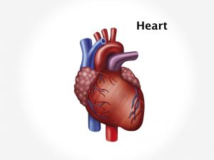
It is a congenital defect and the cause is not known. The tricuspid valve is normally made of three leaflets or flaps. The function of flaps is to allow blood to move from the right atrium to the right ventricle while the heart relaxes and to prevent back flow while the heart pumps. In Ebstein’s anomaly one or two of the flaps may stick to the heart wall and cannot move. It causes leakage of blood from right atrium to right ventricle. In some cases, the flaps are often larger than normal. Sometimes the flaps are in the wrong position in the bottom heart chamber. It is placed too far in the right ventricle. As result of which the right ventricle is smaller than the normal. This causes backflow of blood from right ventricle to right atrium because the leaflets are deep into the right ventricle which prevents outflow of blood from right ventricle to the lungs. In some cases, patients also have a hole in the wall separating the heart’s two upper chambers [ASD-atrial septal defect]. This causes mixing of oxygenated and deoxygenated blood. Thus causing more blood to flow to the lungs which increases work load on heart. Several genetic and environmental factors are responsible for this CHD such as Down’s syndrome in the baby, Rubella or another viral illness during early pregnancy, family history of congenital heart defect. Risk factors during pregnancy include drinking alcohol, poorly controlled diabetes, and certain medications.
Clinical presentation
Depending upon the severity of the complaint complaints may develop soon after birth or in teenage. The baby has laboured feeding, tires easily, has shortness of breath, has heavy or rapid breathing, cyanosis, and frequent pneumonia. Child has poor appetite and no weight gain. Murmur is heard on auscultation of chest. Palpitations in heart , irregular heart rhythm is observed. If there is ASD, oxygenated blood mixes with deoxygenated blood and is transported to lungs. It increases pulmonary blood flow causing lung congestion and symptoms of pulmonary hypertension such as cough, dyspnoea, chest pain leading to congestive cardiac failure. Symptoms of CCF include fatigue, wheezing, excessive sweating, swelling in the legs, sudden weight gain from fluid retention, decreased consciousness.
Investigation
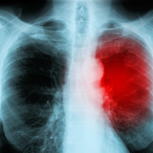
Medical history by the patient’s parents and Clinical examination by the doctor helps in diagnosis. A cardiac murmur can be easily heard on a stethoscope on auscultation which will diagnose CHD. Electrocardiogram [ECG], echocardiogram is recommended. Imaging studies such as Chest x-ray, Magnetic resonance imaging [MRI] of the heart are useful for further evaluation. Cardiac catheterization is also advised in some cases
Treatment
Careful monitoring of the patient is needed if there is no sign and symptoms. Medication to maintain normal heart rhythm and rate is required. Surgical treatment involves only surgical correction of the defective heart. Surgery is needed to close the holes between the heart chambers, and build new tricuspid valve. Avoidance of physical exertion in children is advised.
Recent updates
Researches have been made in surgical robotics and ultrasound guided intracardiac surgery and tissue engineering to stimulate the growth of new tissue to repair congenital defects.
