Causes and risk factors
The causes of ophthalmoparesis include genetic disorders, head injury, stroke, tumurs in the brain, infections, multiple sclerosis, myasthenia gravis, thyroid disorders, migraine, and deficiency of thiamine.
Clinical presentation
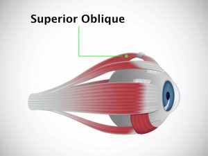
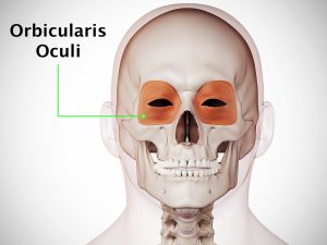
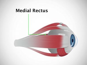
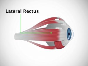
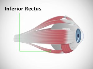
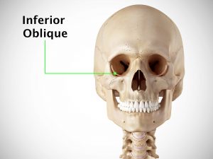
The eye muscles begin to weaken and the eyelids may droop. Patients with ophthalmoparesis have a hard time keeping their eye in a normal position. Even eye movements are very difficult. They may suffer from blurring of vision, diplopia (double vision), ptosis, and dysphagia (difficulty in swallowing). The peripheral vision may be lost.
Investigations
A complete ophthalmoscopic examination is carried out. If necessary, a CT or MRI scan is performed.
Treatment
Treatment for ophthalmoparesis consists of identifying and treating the underlying cause. Spectacles or eye patches can be used to help improve the vision.
Complications
Ophthalmoparesis can worsen to give rise to ophthalmoplegia.
When to contact a doctor
Contact a doctor as soon as you experience any difficulty in eye movement.
Systems involved
Ophthalmology, CNS
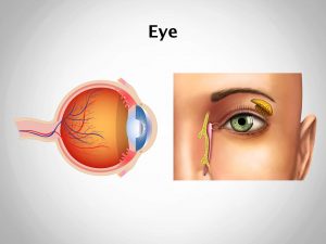
Organs involved
Eyes, brain