Causes and risk factors
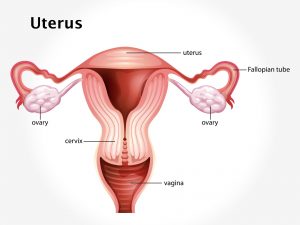
The condition is usually seen in newborns and pregnant women. In newborns, it is seen in premature baby due to improper development and lack of attachment to midline. During pregnancy as the uterus grows, it stretches the abdominal muscle and may cause diastasis recti. It is common during second and third trimester. Multiple births or repeated pregnancies increase the risk of the disease condition.
Clinical presentation
Patient presents with a ridge running in the midline of abdomen from xiphisternum to umbilicus. It is seen prominently during straining of abdominal muscles and disappears when relaxed. Chronic lower back pain due to weakness of abdominal muscle is experienced. In infants it is seen as a bubble under the skin of belly. Extra skin and soft tissue in the front of the abdominal wall may be the only sign of this condition in early pregnancy. In the later part of pregnancy, the top of the pregnant uterus is seen bulging out of the abdominal wall. An outline of parts of the unborn baby may be seen in some severe cases.
Investigation
Medical history by the patient and clinical examination by the doctor helps in diagnosis. Abdominal ultrasound examination is done.
Treatment
For pregnant women, treatment is not necessary. After delivery if the abdominal bulge is affecting the day-to-day activities, then surgery may be recommended. It is also advised for cosmetic purpose. In children, treatment is required in case of complications like umbilical hernia, which then can be treated with surgery.
