Causes and risk factors
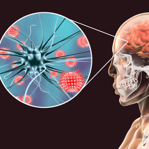
The exact cause is unknown. Factors which increase risk are lack of folic acid in diet of mother during pregnancy, genetic predisposition, exposure to toxins, and infections during early fetal life.
Clinical presentation
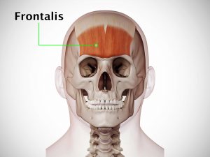
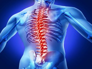
A part of the baby’s brain protrudes out through opening in the skull. It is a sac-like protrusion. Locations of encephalocele include base of skull, areas of nose, sinuses, forehead, or from top of skull to back of skull at the midline. There is a fluid buildup in the brain in patients of encephalocele. Complete loss of strength in the arms and legs is experienced. Uncoordinated movements of voluntary muscles are seen such as those involved in walking, etc. Microcephaly, i.e., unusually small head is observed. There is spastic quadriplegia. Seizures occur. Retarded development is evident. Vision problems, mental and growth retardation, learning difficulties are seen.
Investigation
Medical history by the patient and clinical examination by the doctor helps in diagnosis. Encephalocele can be easily seen during birth, but if it is in the nose, forehead, or sinuses, it can remain unnoticed which can later be diagnosed based upon the signs and symptoms.
Treatment
Surgery is the only treatment. During surgery, the bulging area is placed back into the skull, sac is removed and the accompanying craniofacial abnormality is corrected. Sometimes, shunts are placed to drain excess cerebrospinal fluid from the brain.
Facts and figures
According to CDC, 1 in 10,000 babies born in USA every year are diagnosed with encephalocele.