Causes and risk factors
Certain factors that risk of developing glaucoma are advanced age over 45 years, positive family history of glaucoma, myopia [near-sightedness], hypermetropia [farsightedness], and diabetes mellitus. History of elevated intraocular pressure, past history of eye injury, use of steroid medications, history of diseases like diabetes mellitus, hypertension are some additional predisposing factors.
Clinical presentation
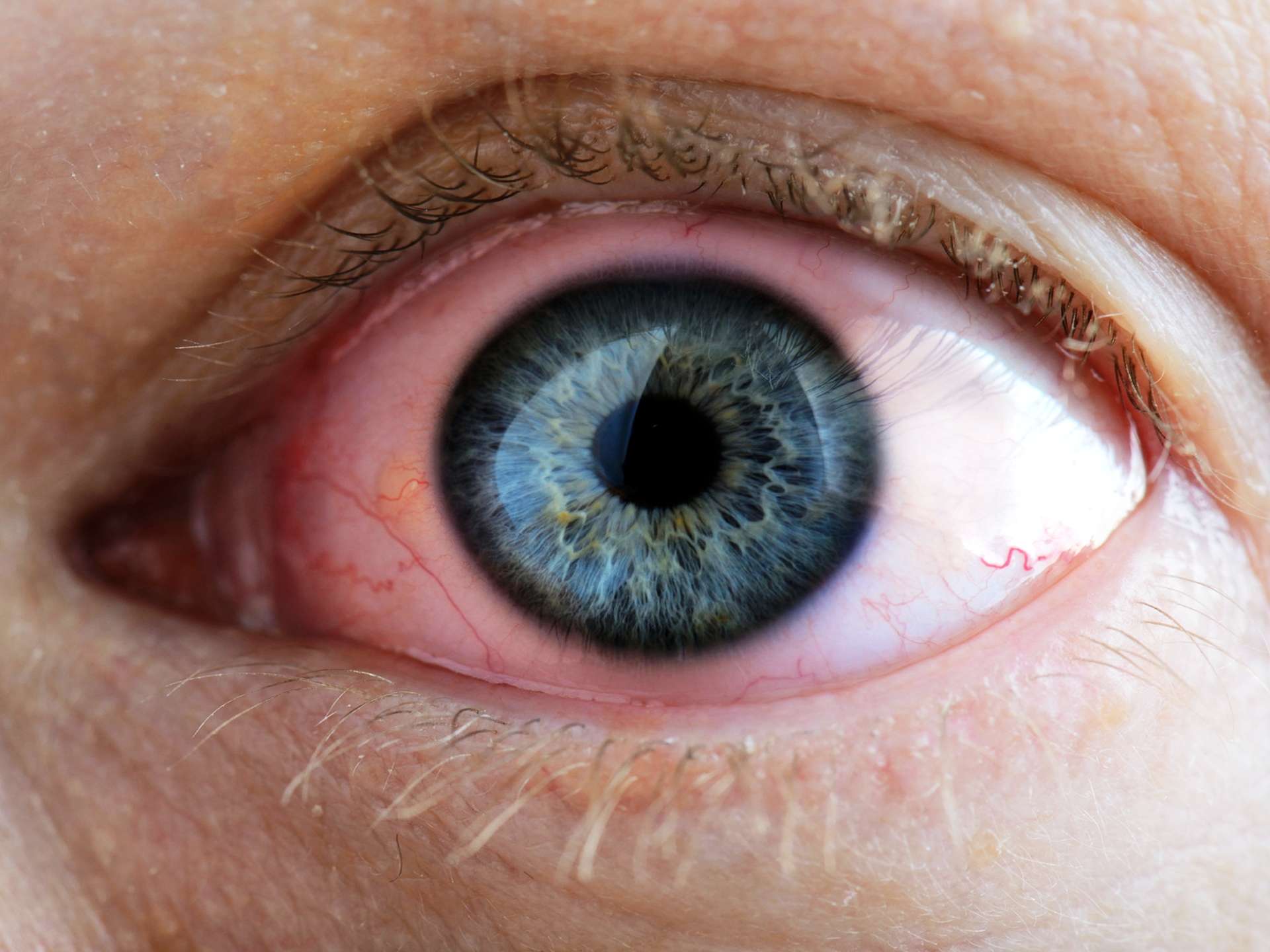
Following are the types of glaucoma – Primary open angle glaucoma in which the drainage angle formed by the cornea and the iris remains open, but fluid in the eye does not flow properly through the drain of the eye, called the trabecular meshwork. This causes fluid to back up in the eye and the intra-ocular pressure gradually rises. This is the most common type of glaucoma. In closed angle glaucoma the drainage angle formed by the cornea and the iris becomes blocked or too narrow due to bulging of the iris. As a result, fluid cannot adequately flow through and exit the eye, causing an abrupt rise in intraocular pressure. In Normal-tension glaucoma the optic nerve becomes damaged even though the eye pressure remains within the normal range. Developmental glaucoma is the one in which the drainage area is not properly developed before birth. A few children may be born with glaucoma or develop glaucoma in the first few years of life. In pigmentary glaucoma, pigment granules from the iris build up in the drainage channels [trabecular meshwork]. Thus the drainage of fluid through the eye becomes slow or completely blocked. Clinical presentation of glaucoma is as follows – many patients may be asymptomatic until the disease progresses. The symptoms include loss of peripheral [side] vision, seeing halos around lights, narrowing of vision [tunnel vision], loss of vision, redness in the eye, hazy eye. Patient complains of pain in eye along with symptoms such as nausea, vomiting.
Investigation
Medical history by the patient and Clinical examination by the ophthalmologist helps in diagnosis. Tonometry in which the pressure within the eye [intraocular pressure] is measured after anaesthetizing the eyes with eye drops is done. Ophthalmoscopy is advised. Visual field testing is recommended. The thickness of the cornea is measured by a test called Pachymeter. Measurement of Visual acuity is done. Gonioscopy is done to inspect the drainage angle and differentiate between open and closed angle glaucoma.
Treatment
Glaucoma cannot be cured and any damage caused by it is irreversible. The treatment is aimed at keeping the intraocular pressure within normal limits to prevent further loss of vision. Eye-drops and oral medications are prescribed. The surgical options are – Laser trabeculoplasty, trabeculectomy in which a small piece of the trabecular meshwork is removed so that fluid can flow through freely from that opening. Drainage implant surgery, Laser peripheral iridotomy is performed in cases of acute closed angle glaucoma. A small hole is made in the iris using a laser so that fluid can flow through it to exit the eye.
Other Modes of treatment
The other modes of treatment can also be effective in treating glaucoma. Homoeopathy is a science which deals with individualization considers a person in a holistic way. This science can be helpful in combating the symptoms. Similarly the ayurvedic system of medicine which uses herbal medicines and synthetic derivates are also found to be effective in treating glaucoma.
