Causative & risk factors
Iodine deficiency in diet is the most important cause of goiter. Although this isn’t encountered frequently in countries where iodized table salt is largely available; it is still a major problem in the mountainous regions of certain countries like India. Intake of cabbage, cauliflower or broccoli in large amounts can also contribute to the development of goiter.
Autoimmune disorders of the thyroid gland such as Hashimoto’s disease and Grave’s disease also cause goiter. A solitary thyroid nodule, thyroiditis or thyroid cancer can also present as goiter. Pregnant women may also develop mild goiter due to effect of human chorionic gonadotrophin (HCG).
Certain kinds of medications and history of radiation therapy to the neck area are also associated with a higher risk of developing goiter.
Clinical presentation
Goiters appear as a visible swelling at the base of the neck. They can be asymptomatic. Sometimes they produce signs and symptoms as a result of pressure on the underlying structures. The patient may experience hoarseness, constriction in the throat and difficulty in swallowing. Coughing or difficulty in breathing may be present.
Depending upon the cause, goiter may be associated with hypothyroidism or hyperthyroidism.
Investigations
A physical exam of the neck is sufficient to detect goiter. Investigations are needed to determine the cause of goiter.
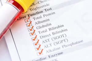
The TSH (thyroid stimulating hormone) level of your blood will be measured along with thyroid hormones like T3 and T4. Blood is also tested for presence of anti TSH antibodies. A thyroid scan is carried out to determine radioactive iodine uptake by thyroid gland.
Your doctor may also suggest an ultrasound, CT or MRI scan of the thyroid gland to visualize it. Occasionally a thyroid biopsy is suggested.
Treatment
In cases where the goiter is small and symptomatic, the patient is only monitored without receiving any actual treatment. Hypothyroidism or hyperthyroidism is treated with thyroid regulating drugs. Oral radioactive iodine is used in some people to shrink the goiter. Wherever these conservative measures fail, surgery is carried out to remove part (partial thyroidectomy) or all of the thyroid gland (total thyroidectomy).
People residing in iodine deficient belts must be encouraged to include adequate quantities of iodized salt must be included in the diet.
Recent updates
Recent studies suggest that fine needle aspiration cytology of the thyroid gland must be done in all cases of simple goiter. Once malignancy is ruled out, goiter must be treated with radioactive iodine uptake or surgery. It has been suggested that the use of levothyroxine (synthetic thyroid hormone replacement) must be stopped completely in cases of simple goiter.


