Causes and risk factors
Two types of HUS are found to be occurring – typical STEC –HUS and atypical – aHUS. HUS is developed due to toxin produced by E coli bacteria in the gastrointestinal system. This is called as shiga like toxin producing E coli HUS i.e. ‘STEC- HUS’. Toxins also destroy RBCs causing kidney injury. Other causes for HUS are salmonella, shigella, campylobacter. Some viruses are also found causing the disease. Atypical HUS or aHUS is a genetic variety of the disorder due to generic defect. It results in complement activation. HUS is common in children. It is most common cause of acute renal failure in children.
Clinical presentation
Hemolytic uremic syndrome is characterized by hemolytic anemia, renal failure [uremia] and thrombocytopenia [low platelet count].Presenting symptoms are diarrhea and vomiting in HUS. The diarrhea is blood stained. There may be hematemesis. Destruction of RBCs in such a way causes hemolytic anemia. Urine frequency is diminished. Associated symptoms include fever, lethargy, weakness and irritability. Later stages shows signs such as bruising on skin, pallor, red spots on skin [petechiae]. Jaundice [yellowness of skin] is observed. There is altered consciousness, seizures. Urine output is reduced.
Investigation
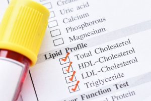
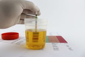
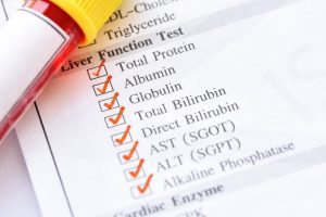
Medical history by the patient and Clinical examination by the doctor helps in diagnosis. Imaging studies such as USG abdomen and pelvis may be useful for the evidence of hepatospleenomegaly or renal abnormalities. CBC for platelets is recommended. Estimation of Prothrombin time [PT] is advised. Comprehensive metabolic panel for BUN, creatinine is done. Urine analysis, urine protein test, renal function test are needed. Stool culture for E coli confirms the diagnosis. Kidney biopsy may be required.
Treatment
Treatment involves hemodialysis. Medications like corticosteroids are administered. Further treatment consists of blood transfusion especially that of packed cell volume [PCV] and platelets.