Causes and risk factors
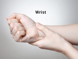
The exact cause of Kienbock’s disease is still not clear. Some studies have put forth certain hypothesis. Some intrinsic and extrinsic factors are responsible for this condition. In most of the people, the lunate is supplied by two blood vessels; however, in a few the blood supply is only by one blood vessel. This causes slow or less blood supply to the lunate bone. Microtrauma due to overuse of the wrist and hand adds up to the causation. Trauma due to fall or severe blow or road accidents can cause injury to the blood supply along with the bone; dislocation or fragmentation of the bone can hamper the blood supply. Pressure on the lunate by ulna during wrist movement is another predisposing factor.
Clinical presentation:
Pain is a prominent feature seen. Along with this, swelling and bruising are the other complaints. Pain is especially seen at the middle of the palm. Movement aggravates the pain. Hence restriction of movement along with stiffness occurs. Weak grip is complained by the patient. On examination, tenderness is present. Apart from these complaints, gradual changes occur in the lunate bone. These changes are divided into 4 stages. In stage I, there are no changes seen and the complaints often mimic wrist sprain. In stage II as the blood supply slows down, the bone starts becoming hard. In stage III, the bone deteriorates further and breaks into pieces, while stage IV is characterized by collapse of the surrounding surfaces of the lunate and arthritis occurs.
Investigations:
Diagnosis is done on the basis of the symptoms narrated by the patient and the physical examination carried out by the orthopedic doctor. Investigations which are done are x-ray of the bone – routine x-rays usually, or specialized digital x-rays along with CT scan and MRI are advised.
Treatment:
There is no complete cure for Kienbock’s disease and hence the aim of the treatment is to restore the blood supply and allay the complaints. The conservative treatment consists of rest and application of ice pack. Analgesic or nonsteroidal anti-inflammatory drugs are advised by the orthopedic doctor for relief of the pain. Cast or splints are recommended for 2-3 weeks. In surgical intervention, various techniques can be adopted. Revascularization for improving the blood supply or leveling of joint is done. In case the lunate is severely collapsed and broken down in pieces, carpectomy is done. Fusion is another surgical technique which can be adopted to relieve the pressure on the lunate.
Other Modes of treatment:
Certain other modes of treatment can also be helpful in coping up with the symptoms. Taking into consideration the symptoms in a holistic way, homoeopathy can offer a good aid for the relief of the symptoms. Certain yoga exercises can also be helpful in strengthening the muscles.


