Causes and risk factors
Activity which involves excessive weightbearing is the main cause of navicular bone fracture. This bone fracture is commonly seen in people engaged in sports like running, jumping, or among soccer players. Athletes and people doing gymnastics or ballet dancers are more prone to such injuries. Studies and researches reveal that military people who indulged in long marches or heavy exercises are highly vulnerable to navicular fractures. Apart from this, certain other factors also contribute to easy fractures; these include poor postures, inadequate diet, muscle weakness, and inappropriate training too. Being overweight causes excessive pressure on the bone, making it susceptible to fractures.
Clinical presentation:
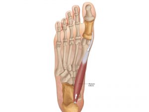
Navicular fractures are of 4 types – Cortical fractures, tuberosity fractures, stress fractures, and fracture of navicular body. Cortical and tuberosity fractures are more commonly associated with ligament injury, while fracture of body of navicula occurs in association with metatarsal joint fracture. Involvement in various sports activity causes stress fractures. Unlike any other fracture, pain is the predominant feature which is aggravated by movement, hence restricted movement is seen. Swelling at the foot, particularly at the mid foot is seen. Movement aggravates pain while rest causes amelioration. Pain radiates to the outer aspect of the foot or to the toe. On examination, tenderness is present. Delay in diagnosis can lead to disability.
Investigations:
Diagnosis is done on the basis of the symptoms narrated by the patient and the physical examination carried out by the orthopedic doctor. Plain x-rays relatively fail for diagnose navicular fracture. Therefore, most of the times these fractures are misdiagnosed. Hence various bone scans are done. Either technetium bone scan, MRI, or CT scan can be done.
Treatment:
Rest and restriction of movement is the first step involved. Analgesic or nonsteroidal anti-inflammatory drugs are advised by the orthopedic doctor. Nonweightbearing cast is advised. Displaced fractures which involve dislocation of the bones need to be corrected by surgical means. For stable fractures, closed reduction is needed. Unstable fractures need to be corrected by internal fixation. Bone growth stimulators which use pulsed electromagnetic fields have been found to be effective. Platelet rich plasma is also used for bone healing.
Other Modes of treatment:
Certain other modes of treatment can also be helpful in coping up with the symptom. Taking into consideration the symptoms in a holistic way, homoeopathy can offer a good aid for the relief of the symptoms. The Ayurvedic system of medicine which uses herbs and synthetic derivates can also be beneficial in combating the complaints. Certain yoga exercises can also be helpful in strengthening the muscles.
