Causes and risk factors
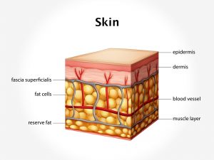
The exact cause of this condition is not known. It is considered to be caused due to certain genetic factors. Mutation of gene GNAQ or GNA11 have been responsible for this condition. Normally, the epidermis consists of the melanocytes which secrete melanin; however, in this condition the melanocytes are seen in dermis.
Clinical presentation:
Appearance of irregular brownish or bluish grey discoloration patch is the characteristic feature seen. The discoloration often resembles as if the area is bruised. It can either be seen on one part of the face or it can affect bilaterally. Usually the discoloration is restricted to the forehead and around the eyes; however, the sclera, retina, and cornea can also be affected. The discoloration can be seen on the oral mucosa. As the age of the child advances, the discoloration tends to become darker and increase in size. The color change can be perceived due to sweating or humidity or during menstruation. Involvement of the eye can lead to glaucoma.
Investigations:
Usually to diagnose these birthmarks, no investigations are required. Clinical examination is sufficient. Skin biopsy and histological examinations can also be done, however, these are rarely advised.
Treatment:
There is no effective treatment available yet for this condition. However, various treatment methods can be adopted. Use of laser therapy or intense pulsed light therapy for destruction of the melanocytes can be done. In cases of involvement of the eyes, the patient is referred to an ophthalmologist. Appropriate conservative and surgical ways of treatment needs to be adopted.

