Causes and risk factors
This disease is not inherited, but an identical heterozygous mutation in the gene PTHR1 has been identified in the affected patients with this disorder. There must be presence of 2 types of genetic mutation for this disease to occur. The development of enchondromas [cartilaginous growth in the bones] could thus be caused by a germline mutation associated with a somatic mosaic mutation.
Clinical presentation
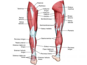
Ollier’s disease occurs bilaterally, but is prominent on one side. Enchondromas develop in the short tubular bones of the hands and feet and long bones of upper and lower extremities. There is development of palpable masses which leads to angular deformity and asymmetrical growth. The masses increase in size as the child grows along with asymmetrical shortening of a limb and either genu varus [outward angulations of the distal segment of a bone] or genu valgus deformities [joint is twisted inwards from the center of the body]. Varus deformity is very common.
Investigations
Medical history by the patient and clinical examination by the doctor helps in diagnosis. Routine blood tests are done. The basic investigation is x-ray of the affected limbs. Biopsy of suspicious lesions is required. CT, MRI, bone scans are recommended. Genetic testing is advised.
Treatment
Treatment involves surgical correction of the deformities. Mechanical aids are helpful for maintaining the physical activities. Physiotherapy, occupational therapy help in managing the condition.
Complications
Complications such as fracture of asymmetrical bones and malignancy can occur.
When to Contact a Doctor
One must consult a doctor if there is growth of the tissues in the limbs
Prevention
Genetic counseling in a couple trying to conceive prevents the disease.
Facts and figures
1 in 100,000 people suffer from this disease.
Systems involved
Genetic system, musculoskeletal system
Organs involved
Bones, cartilages
