Causative & risk factors
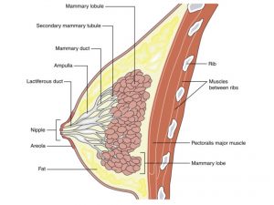
Like most cancers, the exact cause of Paget’s disease of the nipple is not known. The cancer may begin within the nipple itself or it may occur due to spread of cancer cells from the underlying breast tissue.
Clinical presentation
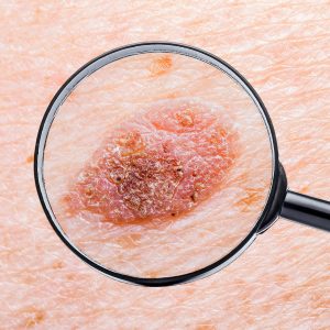
In its initial stages, Paget’s disease of the nipple is only observed as redness and scaling of the skin around the nipple. As the condition advances, there is progressive destruction of the skin. The skin around the nipple appears peeled, thick and with crust formation. Eventually the nipple appears flattened against the breast tissue. The patient may complain of severe itching, tingling, tenderness and burning pain in and around the nipple.
Since this condition is frequently associated with breast cancer, a lump is usually felt on breast examination. Discharge from the nipple may be present, which is usually bloody in character.
This condition usually occurs in only 1 nipple, although rarely both nipples may be involved. Even though Paget’s disease begins initially in the nipple, it eventually spreads to involve the areola and sometimes other regions of the breast.
Investigations
First a local examination of the breast is performed. Any nipple discharge if present is sent for examination. Then a biopsy may be carried out to detect underlying malignancy. Imaging studies such as a mammogram or MRI scan are usually suggested.
Treatment
Since Paget’s disease of the nipple is frequently associated with breast cancer, mastectomy i.e. removal of the breast tissue along with the lining of the chest muscles is the treatment of choice. If the malignancy is extensive, the axillary lymph nodes are also removed; this procedure is known as radical mastectomy.
On the other hand, if the malignancy is confined to the nipple, only a part of the breast tissue is removed, whilst conserving the remaining breast tissue (lumpectomy).
Following surgery, radiation therapy or chemotherapy may be suggested to prevent recurrence of malignancy.


