Causes and risk factors
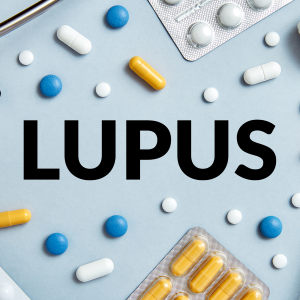
Pericardial effusion can be caused by a variety of reasons. Pericarditis (inflammation of the pericardium) is the most common cause of pericardial effusion. Infections either bacterial, viral or add up to the causation. Pericardial effusion can be caused due to certain underlying heart diseases like myocardial infarction, autoimmune diseases like lupus, hypothyroidism or end stage renal disease. Cardiac surgery can also lead to pericardial effusion. Pericarditis can be caused due to any trauma or injury to the heart. Cancer spread of cancer or the radiation and chemotherapy used during cancer treatment also lead to pericardial effusion.
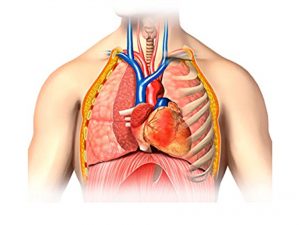
Clinical presentation
Usually the patient does not suffer from any complaints. Symptoms appear only when large amount of fluid is accumulated. This accumulation of fluid can lead to compression on the heart and its surrounding structures. The patient comes up with complaints of pain and feeling of fullness and heaviness in chest. Fullness is felt in abdomen. Nausea, difficulty in breathing and difficulty in swallowing are the other associated complaints seen. The mental status of the person can also be altered. On examination bluish discoloration of skin and nails is seen. The excessive accumulation of fluid causes compression on heart and can lead to disturbance in the functioning of the heart. This condition of heart is cardiac tamponade. It is one of the serious complications of pericardial effusion. Shock is another common complication seen.
Investigations:
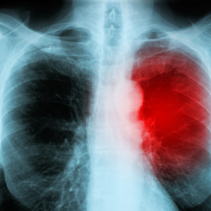
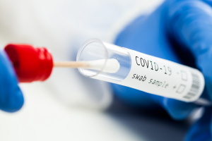
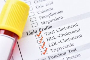
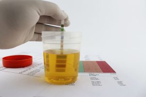
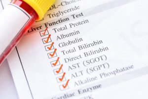
Diagnosis is done on the basis of the symptoms narrated by the patient and the physical examination. The required investigations are chest X ray, ECG and CT scan of the chest.Pericardiocentesis where the fluid is extracted for examining is done.Certian other investigations like routine blood test, urine test, renal function test, liver function test, blood sugar levels etc can also be done.
Treatment
The treatment depends upon the cause. Medications like non steroidal anti inflammatory drugs, antibiotics or diuretics are started. Draining of the excessive fluid is the main line of treatment.Ultrsaound guided pericardiocentesis is done for this. Video assisted thoracoscopic surgery is another non invasive technique available for draining of the excessive fluid. Cases which cannot be managed by these non invasive technique requires surgically managed. Percutaneous balloon pericardiotomy or n Subxyphoid Pericardiostomy can be done.
Other modes of treatment:
Certain other modes of treatment can also be helpful in coping up the symptom. Taking into consideration the symptoms in holistic way, homoeopathy can offer a good aid for the relief of the symptoms. The Ayurvedic system of medicine which uses herbs and synthetic derivates can also be beneficial in combating the complaints.