Causative & risk factors
A viral (influenza), bacterial (tuberculosis or pneumonia) or fungal infection can give rise to pleurisy. Pleurisy can also result from rheumatic diseases (SLE, RA), cancer of the lung and intake of certain medications. Trauma to the chest can also cause pleurisy.
Clinical presentation
A patient with pleurisy experiences severe, sharp pain usually on 1 side of the chest. This pain is greatly exacerbated on deep breathing, coughing, sneezing or talking. This pain disappears on holding the breath. Since deep breathing is painful, the patient may start taking shallow breaths in rapid succession. The chest pain may radiate to other locations such as the abdomen, shoulders or neck area.
Sometimes, extra fluid can build up into the pleural space. This is known as pleural effusion. Pleural effusion may produce symptoms like shortness of breath and coughing. Pleural effusion must be promptly treated by your health care provider.
Investigations
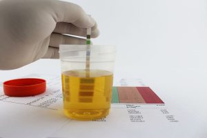
Investigations are carried out primarily to detect the cause of pleurisy. Blood tests are carried out to detect evidence of infections. Imaging tests such as Chest X ray, ultrasound, CT or MRI scan are carried out to visualize the lungs clearly.
A procedure known as thoracocentesis is carried out to extract and analyze the pleural fluid. Biopsy of the outer membrane of the pleura can be carried out in order to rule out cancer.
Treatment
Any underlying infection is treated with antibiotics, anti virals or anti fungals depending upon the causative organism.
Painkillers and anti-inflammatory drugs like NSAID’s are prescribed to lessen the pain and discomfort. If too much fluid has accumulated in the pleural space, thoracocentesis is carried out to extract the fluid.
Recent updates
A new test known as Xpert MTB/RIF assay has been developed for diagnosis of pleural tuberculosis. The pleural tissue or pleural fluid can be used to carry out this test.