Causes and risk factors:
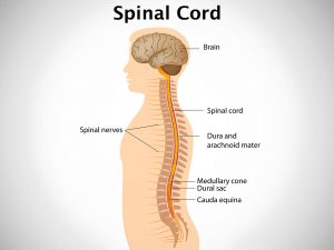
The spinal cord developes from the neural plate. The development begins in early pregnancy and it is the last to complete with its development. This neural plate forms a groove which further undergoes changes to form a tube (neural tube) like structure. The top of the tube forms the brain and the rest becomes the spinal cord. Failure of this process results in various developmental defects of brain and spinal cord. It comprises of spina bifida also. The exact cause of spina bifida is not known. Certain genetic factors are the predisposing causes.
Clinical presentations:
There are four main types of spin bifida- spina bifida occulta and spina bifida aperta. Meningocele and myelomeningocele. Some patients can remain a symptomatic, while some presents with complaints. The symptoms of spina bifida vary from person to person and as per the type. The child can present with a dimple in the skin at the region of the spinal cord,a soft mass can be felt on palpation or a dermal sinus or tuft of hair can be seen at the lumbosacral region.As the muscles is damaged and the nerve supply is hampered foot deformities can be seen. A fluid filled sac can be seen protruding on the back or at time an exp abnormal developed spinal cord tissue without any skin covering is seen. Mild to severe paralysis of the limb and contracture of the hip, bowel and urinary incontinence are the other symptoms seen.
Diagnosis and investigations:
Diagnosis can be done wither during pregnancy or after birth. Pre natal checks ups will help to evaluate the spinal development. Ante natal USG anomaly scans in 18-20 weeks, amniocentesis aid in diagnosis. Blood test for AFP levels and genetic markers in mother help in detection before birth. In some cases the defect is not diagnosed until the baby is born. The symptoms narrated by the mother/ partents and examination of the baby can help in diagnosis. X-ray, CT scan or MRI can be advised to get a clear view.
Treatment:
There is no specific cure for spina bifida. Asymptomatic patients do not require any treatment. While some cases where aggravation of complaints are seen require surgical intervention. Surgical intervention is required to close the defect and to correct the deformities. Although experimental fetal surgery in uetro is performed which helps to correct the defect earlyUse of assistive devices like braces, crutchers, walkers, wheelchairs can be advised.Special exercises are started at an early age. The bowel and bladder incontinence is treated with medications or by catheterizations. Intake of folic acid under prescription during pregnancy is the recommended preventive measure.
Recent Update:
The National Institute of Neurological Disorders and Stroke (NINDS), a part of the National Institutes of Health (NIH) is conducting researches for looking at the hereditary basis of neural tube defects. It is working to identify, characterize, and evaluate genes for neural tube defects so as to understand the genetics of neural tube closure, and to develop information that will help for treatment planning and genetic counseling.
Studies that shed light on how folic acid prevents spina bifida and may lead to improved forms of folate supplements are in progress.