Causes and risk factors
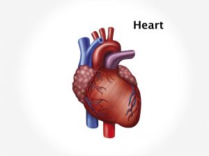
No particular cause is known.VSD is as a developmental abnormality due to genetic problem. The septum is made up of inferior muscular and superior membranous portion. During foetal development the septum fails to form fully to separate right and left side of heart between lower chamber of heart causing VSD. 4 types of VSD are known – concoventricular i.e. hole appears in the septum near aortic and pulmonary valves, perimembranous – upper section of septum, inlet ventricular VSD – in the septum near mitral and tricuspid valves or muscular VSD – lower part of septum. Membranous VSD is the most common type. It can be hereditary problem. It may be present along with other heart abnormalities in many children. Children with Down’s syndrome are prone to have VSD.
Clinical presentation
Symptoms depend upon size of the hole. Symptoms like Murmur is heard on auscultation of chest. Smaller the hole louder the murmur. Small holes generally cause little or no difficulty. They close usually upto 2 years of age. A baby with larger ventricular septal defect shows laboured feeding, tires easily, has shortness of breath, has heavy or rapid breathing, poor appetite, and no weight gain. Left ventricle carries oxygenated blood while right ventricle carries deoxygenated blood. VSD allows shunting of blood from one ventricle to the other. When a VSD allows shunting of blood from left to right ventricle i.e. oxygenated blood mixes with deoxygenated blood and is transported to lungs. It increases pulmonary blood flow causing lung congestion and symptoms of pulmonary hypertension such as cough, dyspnoea, chest pain leading to congestive cardiac failure. When a VSD allows shunting of blood from right to left i.e. deoxygenated blood mixes with oxygenated blood and is transported to whole body. Impure blood reaches all the tissues and organs thus depriving them of oxygen.
Investigation
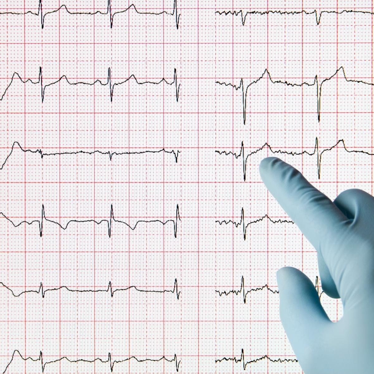
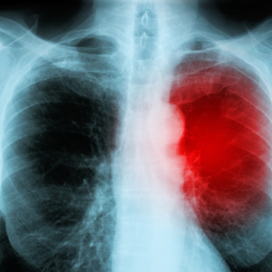
Medical history by the patient and Clinical examination by the paediatrician helps in diagnosis. A cardiac murmur can be easily heard on a stethoscope on auscultation which will diagnose VSD. Pulse oximetry measures amount of oxygen% in the blood. A chest X ray, ECG, Echocardiography, cardiac catherization will confirm the diagnosis.
Treatment
Treatment depends upon the size of the opening in septum. Small openings in the ventricular septum close on their own and do not need any treatment but the baby is observed until the defect is cleared. Larger openings are closed by open heart surgeries. Avoidance of physical exertion in children is advised for larger VSD. Medications that help in case of VSD are those which increase the heart contractions, reduce the blood volume and regularise heartbeats. Poorly fed babies [due to exhaustion while feeding] require high calorie diet or use of feeding tube.
Recent updates
Researches have been made in surgical robotics and ultrasound guided intracardiac surgery and tissue engineering to stimulate the growth of new tissue to repair congenital defects.
